Pioneers in Molecular Imaging and Theronostics
Cardiac PET/CT
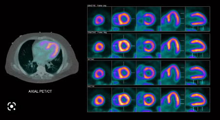
Cardiac PET/CT is one of most accurate ways to diagnose coronary artery disease and detect areas of low blood flow in the heart. It requires the availability of a special radioactive tracer produced on-site, and CIRA is the first private center in Florida to install a medical cyclotron and produce N-13 Ammonia on-demand. With new state-of-the-art technology and dedicated personnel, we can accurately detect the presence of CAD and provide precise quantitative measurement of blood flow to heart muscle.
Oncology PET/CT
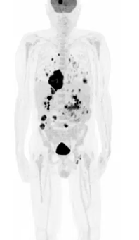
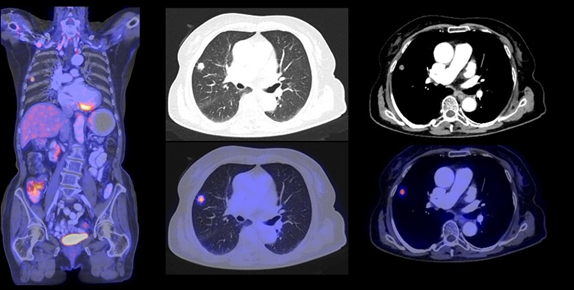
F-18 FDG PET/CT is a powerful imaging technique in the diagnosis, staging, and re-staging of cancer. It relies on the fact that cancer cells typically have a higher rate of glucose metabolism than normal cells. This technology allows for highly detailed images that help in several ways:
- Diagnosis: F-18FDG PET/CT can identify the presence of cancer in the body by pinpointing areas with increased glucose uptake, enabling the detection of even small or hidden tumors.
- Staging: For patients with known cancer, it is used to determine the extent and spread of the disease, helping physicians to plan the most appropriate treatment approach.
- Re-Staging: In cases where cancer has recurred or is suspected to have metastasized, F-18FDG PET/CT can help identify new tumor locations, assess the effectiveness of prior treatment, and guide subsequent therapeutic decisions.
F-18 FDG PET/CT is a valuable tool in oncology, offering precise information that aids in diagnosis and in the management of cancer patients throughout their treatment journey.
PSMA PET/CT
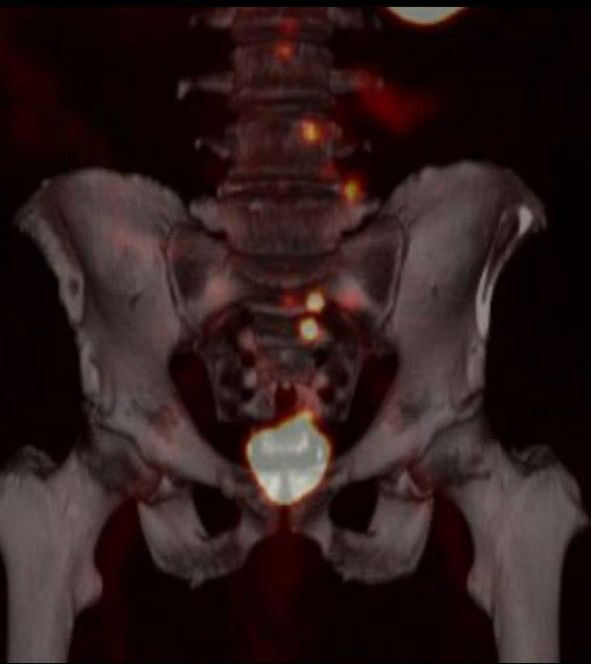
PSMA imaging is a breakthrough in prostate cancer care due to its precision, early detection capabilities, and its role in individualized treatment and monitoring. It can significantly improve the way prostate cancer is diagnosed and managed, and CIRA Health offers this important Molecular Imaging study.
PSMA (Prostate-specific membrane antigen) imaging is new and helpful for prostate cancer for a few key reasons:
1. Precision: PSMA scans offer highly precise images that can detect even very small amounts of prostate cancer cells. This level of accuracy was not previously possible with conventional imaging techniques.
2. Early Detection: The sensitivity of PSMA imaging allows for the early detection of prostate cancer and its metastasis, enabling treatment at earlier and potentially more curable stages.
3. Treatment Planning: It provides detailed information about the location and extent of prostate cancer, aiding doctors in planning personalized treatment strategies. This helps in tailoring therapies to the patient's specific needs.
4. Monitoring Response: PSMA scans are also used to monitor how well a patient is responding to treatment, which is crucial for adjusting the treatment plan as needed.
5. Reduced Invasiveness: It is a non-invasive procedure, meaning there's no need for biopsies or exploratory surgeries to locate cancer cells, reducing patient discomfort and risk.

NEUROLOGY
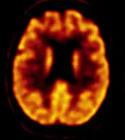
Amyloid PET/CT scans are valuable in the diagnosis of Alzheimer's disease because they can detect the presence of abnormal protein deposits known as amyloid plaques in the brain. Amyloid plaques are a hallmark of Alzheimer's disease and are often associated with cognitive decline. Amyloid PET scans are used in Alzheimer's disease diagnosis:
- Early Detection: Amyloid PET scans can detect amyloid plaques in the brain before significant cognitive symptoms appear. This early detection is crucial for diagnosing Alzheimer's disease in its early stages when interventions may be more effective.
- Accurate Diagnosis: These scans provide more accurate and definitive confirmation of Alzheimer's disease, helping to differentiate it from other forms of dementia or cognitive disorders with similar symptoms.
- Tailored Treatment: Knowing the presence of amyloid plaques can aid in the development of individualized treatment plans and the monitoring of disease progression.
The approval of two new drugs for the treatment of Alzheimer’s disease is a significant development as they target amyloid plaques in the brain to slow progression of the disease.
Ultrasound
Ultrasound is a quick and easy exam that is often used as a first diagnostic imaging step.
Diagnostic CT
CT combines the power of X-rays with computers to capture multiple pictures of the inside of your body. CIRA offers all diagnostic CT studies including Cardiac Calcium Screening and Lung Screening.
SPECT/CT
A SPECT/CT is a combination of a SPECT (Single Photon Emission Computed Tomography) scan with a CT (Computed Tomography) scan. A SPECT scan is a type of nuclear medicine test that uses a radiotracer (a special contrast agent), that is injected through your vein. A CT scan uses X-ray radiation to provide thorough images of the structures inside your body (anatomy).
By integrating both of these exams, the technology creates a very detailed and informative study by showing both your anatomy and physiology. The combination of both the SPECT and CT are used to help avoid, detect and treat an assortment of abnormalities within the body. Many times a SPECT/CT can identify the disease, even at its early stages before other imaging exams.
- CIRA MIAMI 2760 SW 97th Ave, Miami, FL 33165, United States of America
- CIRA SARASOTA 2000 S Tamiami Trl, Sarasota, FL 34239, United States of America
- CIRA MIAMI BEACH 300 Arthur Godfrey Rd, Miami Beach, FL 33140, United States of America
- CIRA KENDALL 9408 SW 87th Ave, Miami, FL 33176, United States of America
BROWSE OUR WEBSITE
CONTACT INFORMATION
Phone: 305.594.2881
Email: info@ciramiami.com
Fax Number: 305.594.2871
Address: 2760 SW 97th Ave. Suite B-101
Miami, FL 33165
- Mon - Fri
- -
- Saturday
- Appointment Only
- Sunday
- Closed
Se Habla Espanol
OUR LOCATIONS
CIRA MIAMI
2760 SW 97 AVE SUITE B-101 MIAMI FL 33165
CIRA SARASOTA
2000 SOUTH TAMIAMI TRAIL
SARASOTA FL 34239
CIRA MIAMI BEACH
300 ARTHUR GODFREY ROAD SUITE # 101
(Coming soon 2024) MIAMI BEACH FL 33140
CIRA KENDALL
9408 SW 87TH AVE SUITE # 104 (Coming soon 2024) MIAMI FL 33176
Phone: 305.594.2881
Email: info@ciramiami.com
Fax Number: 305.594.2871
Address: 2760 SW 97th Ave. Suite B-101
Miami, FL 33165
